
• Case Provider: Dr. Zheng Liu
• Solution Used: Eighteeth Helios 500 Intraoral scanner and FinScan F350 CBCT
• The treatment plan includes obtaining 3D data through intraoral scanning, designing a surgical guide for a restoration-oriented implant procedure, and placing the upper structure during the second-stage restoration aftel the implant. A non-cemented retention restoration solution will be used. The intraoral scanning service team and the implant team will provide clinical technical support.
• About the Dentist
Dr. Zheng Liu graduated from the College of Stomatology at Shanghai jiao Tong University. in 1999, he pursued further studies in Japan through a Sino-Japanese friendship Program, and in 2006, he was appointed as a guest lecturer at the College of Stomatology at Shanghai jiao Tong University and Shanghai University of Medicine and Health Sciences.
• Case
The patient is a 43-year-old male with a missing tooth at position 46 for over a year.
He is in good health with no history of allergies.
• Treatment Process
The preoperative intraoral 3D model of the patient was obtained using the Helios 500 intraoral scanner.

Bone tissue information of the patient was obtained using Eighteeth's FinScan F350 CBcT. The CBcT data was reconstructed and aligned with the intraoral scan model data in the design software, while simultaneously determining the position of the mandibular nerve canal.

Outline the position and course of the mandibular nerve canal in the design software.

The preliminary tooth arrangement is used as a guide for implant planning.

By reconstructing the 3D digital model, the implant position and angle are planned with a restoration-oriented approach. This strategy minimizes surgical risks while reducing lateral shear forces on the implant, ensuring that the transmission of occlusal forces aligns more closely with biomechanical principles.

Design the surgical implant guide and proceed with 3D printing.


By using Eighteeth's FinScan F350 CBCT and Helios 500 intraoral scan data, confirm the implant insertion angle and site, as well as the bone density around the implant. This information is essential for determining the surgical plan and driling sequence.

Two months postoperatively, the follow-up shows that the implant site aligns with the design plan.

It can be observed that the implant angle is consistent with the design plan.

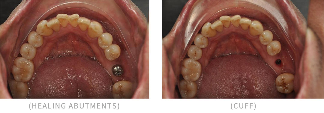
Remove the healing abutment and use the Helios 500 intraoral scanner to perform an intraoral scan, capturing the soft tissue emergence profile and marking the tooth positions.

Install the scan body intraorally. Take a radiograph to confirm the position of the scan body.

Use the Helios 500 intraoral scanner to obtain the scan bodyinformation.

Scan the occlusal relationship.

Align the database with the scan body. Generate the digital prosthesis.

Design the final restoration.

3D print the gingiva and the model. Place the final restoration crown on the model to check occlusion and interproximal contacts.

Use the non-cemented abutment technigue to install the restoration crown, avoiding any residual adhesive.

Photo of the fitted crown

• Conclusion
In recent years, digital technology has been driving continuous advancements in the field of dentistry, greatly facilitating the clinical work of practitioners and bringing more benefits to patients. Choosing a good digital tool can significantly enhance the efficiency of doctors' work.
In this case, the patient sufered from chronic pharyngitis, which made conventional silicone rubber impressions dificult due to severe gag relex. However, the Helios 500 intraoral scanner allowed for a smooth and comfortable impression process, leaving the patient very satisfied.
The Helios 500 intraoral scanner saved doctors a considerable amount of time and reduced cumulative errors associated with the tedious steps of traditional gypsum model fabricaion. This enhanced the predictability of the final restoration and improved the accuracy and speed of data acquisition for clinicians.